Arthroscopic Knee Surgery

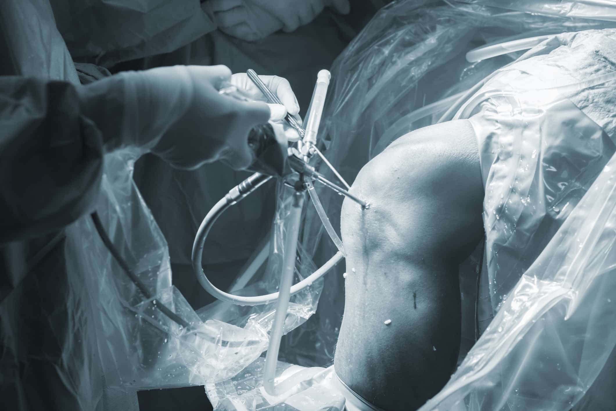
Arthroscopy is a minimally invasive procedure that allows doctors to examine tissues inside the knee. During an arthroscopic procedure, a device known as an arthroscope is inserted into a small incision in the knee. Through this tube, a thin fiberoptic light, magnifying lens and tiny video camera are inserted, allowing the doctor to examine the joint in great detail. Arthroscopy may be a diagnostic procedure following a physical examination and imaging tests such as MRI or CT scans or X-rays. It may also be used as a method of treatment to repair small injuries in the knee.
Knee Arthroscopy as Treatment
Relatively minor knee damage is frequently treated using arthroscopic techniques. Most knee damage results from sports injuries or osteoarthritis. During an arthroscopic procedure, the surgeon may be able to treat:
- Loose bone or cartilage
- Meniscal tears
- Torn ligaments
- Synovitis (swelling of the joint lining)
- Misalignment of the patella (knee cap)
- Inflamed tissue
In patients with osteoarthritis of the knee, arthroscopy is also used in the removal of dead tissue, a process known as debridement.
Benefits of Knee Arthroscopy
Because it is minimally invasive, arthroscopy offers the patient many advantages over traditional, more invasive, surgery. These include:
- No cutting of muscles or tendons
- Smaller incisions
- Less bleeding during surgery
- Less scarring
- Shorter recovery time
- Shorter and more comfortable rehabilitation
Candidates for Knee Arthroscopy

Knee arthroscopy is quickly becoming the ideal procedure for many conditions affecting the knee. Its minimally invasive advantages allow patients to receive fast and simple pain relief, increased range of motion and restored function, while avoiding or delaying the need for joint replacement surgery. Despite its many advantages, arthroscopy is not appropriate for every patient. Some patients, especially those with knee problems that are in difficult-to-see areas, may benefit more from conventional surgery.
The Knee Arthroscopy Procedure
Knee arthroscopy is performed on an outpatient basis under local or general anesthesia, depending on the type and severity of the condition, as well as the patient’s personal preference. During the procedure, the surgeon inserts the arthroscope into the knee through a tiny incision. This instrument is used to identify any damage or abnormalities within the knee, or to confirm the diagnosis of a previous imaging exam.

If damaged areas are detected, they can be repaired during the same procedure by inserting surgical instruments into additional small incisions.
Recovery from Knee Arthroscopy
After a knee arthroscopy , patients often experience swelling and pain for several days. These symptoms can be controlled by the usual home remedies: resting and elevating the leg, applying ice and taking over-the-counter painkillers. Patients are encouraged to get up and walk around as soon as possible after the procedure, although crutches or a cane may be needed for some period of time.
Most patients can usually return to work within a week, but will need to undergo physical therapy in order to restore full range of motion to the joint. Most patients can resume light physical activities after a few weeks, although full recovery from knee arthroscopy may take 12 weeks or longer.
See What Our Patients are Saying:
Risks of Knee Arthroscopy
While knee arthroscopy is considered safe for most patients, there are certain risks associated with any surgical procedure. These risks include: infection, blood clots, accumulation of blood in the knee, nerve damage or adverse reactions to medications or anesthesia. In the great majority of cases, the knee arthroscopy goes smoothly.
Posterior Cruciate Ligament Tears

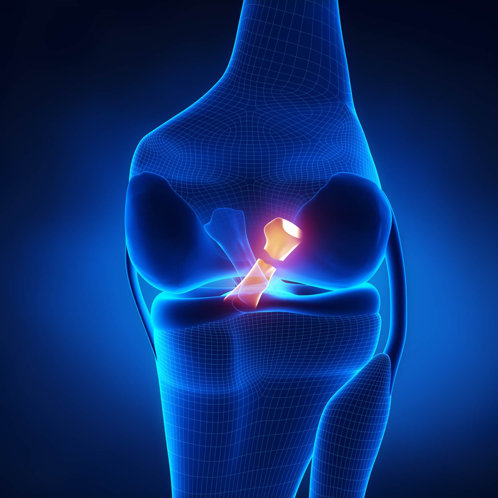
The posterior cruciate ligament (PCL) is one of four ligaments that helps support the knee and protects the shin bone (tibia) from sliding too far backwards. The cruciate ligaments are located inside the knee joint and cross over each other, forming an “X”. The anterior cruciate ligament is in the front and the posterior cruciate ligament is located behind it in the back of the knee. These ligaments control the back and forth motion of the knee.
Injury to the PCL most commonly occurs when the knee is bent and an object strikes the shin, pushing it backwards.
This is commonly referred to as a “dashboard injury” because it often happens during a car accident when the shin is forcefully pushed into the dashboard. A PCL tear may also be caused by a sports injury or a fall. In many cases, a posterior cruciate ligament tear occurs along with injuries to other parts of the knee, including other ligaments, cartilage and bone.
Symptoms of a PCL Tear
Individuals with PCL tears may experience pain,swelling and limited range of motion within the knee. Some people may also experience a feeling that the knee has popped or given out, as it causes instability within the joint. In most cases, a PCL tear will make it difficult to walk.
Diagnosis of a PCL Tear
A PCL tear is diagnosed through a physical examination of the knee. Additional imaging tests may include an X-ray or an MRI scan which can show clearer images of a posterior cruciate ligament tear and help to determine if other knee ligaments or cartilage are also injured. A diagnostic arthroscopy may also be performed to view detailed images of the tear.
Treatment of a PCL Tear
Treatment for a PCL tear varies based on the severity of the injury, but it is commonly treated with conservative methods that may include:
- Rest
- Ice
- Gentle compression
- Elevation
- Immobilization with a knee brace
A physical therapy program may help to strengthen and restore function to the knee. If there is significant swelling that interferes with the mobility of the knee, a joint aspiration procedure may be performed to drain any excess fluid from the joint. In severe cases, when the ligament has torn completely and not healed properly, surgery may be necessary to repair or rebuild the ligament.


